Friday, April 27, 2012

West Nile Virus: A Case Study


West Nile Virus- A Case Study

 By: Shannon Hinton

Signalment/History
It the middle of July and you get a call from an owner that says her horse, ‘Lucky’- a 5 year old Thoroughbred mare, seems to be dropping a lot of feed. She also says the horse is acting depressed and doesn’t seem to be walking well. The owner says that these problems appeared earlier yesterday morning. The horse has not had any previous medical issues. When asked about vaccination status, the owner said that she ‘did not believe in giving her horses shots.'
Horse showing ataxia and incoordination. Photo from:
ckequinehospital.com


Physical exam
You go out to the farm to look at the horse. Upon arrival, you notice buckets of standing water and presence of mosquitoes. Upon examination, you note: 
  • Fever- 102 F
  • Incoordination
  • Ataxia of the hindlimbs 
  • Lethargy 
  • Dysphagia 
  • Muscle fasiculations of the muzzle 
Differential Diagnosis
  • West Nile Virus 
  • Equine Protozoal Myeloencephalitis
  • Equine Herpes Virus 1 
  • Rabies 
  • WEE, EEE, VEE  
Rabies: You would expect to see more cerebral involvement consisting of behavioral alterations, depression, seizures and coma. Horses with rabies are also prone to regurgitation through the nose.  Also, no wounds were seen on the horse, so she was probably not bitten by a rabid animal. Mortality in unvaccinated horses is 100%.
EPM: Can be difficult to diagnose, but the presence of a fever and muscle fasciculations of the face point towards West Nile Virus over EPM.
EEE/WEE: More CNS signs are expected including, drooping of the head, chewing, head pressing, circling, convulsions and excessive salivation. A biphasic course with fever, remission, then fever again with CNS signs is also common. Mortality of unvaccinated horses infected with EEE is also quite high, so this horse would not be expected live.
EHV-1: Hindlimb to whole body paresis is to be expected in an EHV-1 infection. Bladder atony and incontinence is also expected. Respiratory signs could also accompany neurological signs in an EHV-1 infection. 

Initial Diagnostic Plan
  • CBC
  • Chemistry 
Results of bloodwork
  • CBC:  leukogram showed a mild inflammation and stress response, dehydration was noted 
  • Chemistry:  hyponatremia was present (WNV is speculated to cause inappropriate release of anti-diuretic hormone)

http://www.inbios.com/elisas/west-nile-detect-igm
Diagnosis

Based on clinical signs, lack of vaccination status and presence of mosquitoes you decide to take a blood sample and send it out to a lab to look for the presence of West Nile Virus using an IgM capture ELISA. Upon receiving the results, you note there is a large rise in IgM antibodies. You know that the IgM capture test will detect antibodies seven days to three months post WNV exposure. These results, clinical signs and lack of vaccination allow you to make the diagnosis of West Nile Virus infection.  
 

http://www.idexx.com/pubwebresources/pdf/en_us/livestock-poultry
/igm-wnv-information-brochure.pdf
 
Treatment

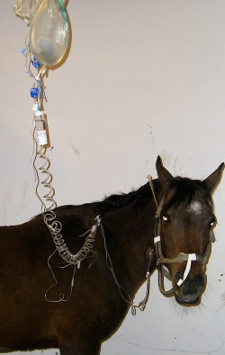
Photo from ckequine.com
Treatment is supportive only, as there is no antiviral therapy for flaviruses. NSAIDs, such a flunixin meglumine, can be administered to help combat muscle fasciculations. Fluids should be administered to replace water and electrolytes losses. The animal should also receive assisted enteral nutrition via a nasogastric tube until swallowing capabilities return.

The owner asks if she should move her other horses so that they do not become infected with WNV. You tell her that that will not be necessary because horse to horse transmission of the virus does not occur. The best means of protecting her other horses is to control the mosquito population around the farm and to vaccinate the remaining horses. The WNV vaccine is an inactivated whole virus vaccine. The vaccination series consists of two vaccinations given 3-6 weeks apart initially and then annually. Ideally the vaccine should be given before the start of mosquito season. It is labeled for prevention of viremia.
 
Follow Up

Lucky was showing marked improvement three days following the initial visit. She has regained her ability to swallow and is able to have the NG tube removed. Her gait and incoordination has also shown improvement, and you believe that she will not have any long lasting effects. Horses that have survived WNV infection are immune for life. 

Resources
1. Reed, Stephen, Warwick Bayly, et al. Equine Internal Medicine. 3rd ed. St. Louis: Saunders, 2010.
2. Sellon, Debra and Maureen Long. Infectious Equine Disease. 1st ed. St. Louis: Saunders, 2007.
3. Subbiah, E. VM 8124 Course Notes, Equine Viruses I. Spring 2012; Lecture 13-15.

Prevention Tests and Treatment of West Nile Virus

Prevention Tests and Treatment of West Nile Virus

By: Katherine Robinson
Prevention of West Nile Virus

            The prevention of West Nile virus infection of people and horses is very similar. The best way to prevent West Nile virus is to prevent the mosquitoes from biting. This can be done in many different ways which include reducing the mosquito population, using insect repellent in many different forms, and being conscious of the hours that they are out and ready to bite. The mosquitoes feed the most at night on hot days so horses in endemic areas should be kept in the barns at night time. All stagnant water should be dumped to prevent a site for mosquitoes to replicate. Frequently removing feces and keeping fans on around horses to keep the air moving will help reduce mosquitoes in the barns. Mosquito repellents that are approved for the horse should be applied to reduce the possibility of bites. In horses, vaccination is the primary method to decrease the risk of infection, but doesn’t fully protect from clinical disease.  Dead birds can indicate WNV is in the area so dead birds should be reported to the local authorities to help monitor for presence of WNV.

            Since risk of exposure varies, and West Nile virus is such a serious disease, it is recommended by the American Association of Equine Practitioners (AAEP), that all horses in North America be vaccinated for the virus. There is an inactivated vaccine and live canarypox vaccine. The vaccine has been shown to prevent viremia in horses for 12 months after the vaccine has been given, but should be given more frequently, every 6 months, in West Nile virus endemic regions. Vaccination is best in the spring before the vector season reaches its prime. Mares should be vaccinated before they are bred because neither of the vaccines is licensed for pregnant mares, although it has become an accepted practice to vaccinate horses while pregnant because the risks of infection outweigh the risks of adverse vaccine effects. However, it should be noted that modified, live vaccines should not be used in pregnant animals. Mares are also vaccinated 4-6 weeks before foaling to pass viral immunity along to their foals through colostrum. Foals should receive a vaccine of their own at 3-4 months, after the colostral immunity has worn off, then again at 1 year.

Testing for West Nile Virus

            Identifying infected horses should be done with an antibody test and should use horse serum or CSF. The horses that are tested are ones who show clinical signs of WNV, as this is not a routine test done on all horses. IgM capture ELISA, or plaque-reduction neutralization are the available tests for veterinarian diagnosis. IgM antibodies are expressed earlier and at a higher concentration then IgG in WNV infected horses. Tests should detect IgM rather then IgG because it is a better sign of an active infection. IgG tests can be done to test for previous infection of WNV or secondary infection. When testing for IgM it is important to test early in infection because the antibodies do start to decrease around 4 days after clinical sign onset. Tissues from dead horses should be analyzed with real time PCR.

West Nile Virus Treatment

            Treatment for horses that are infected is supportive, like treatment for most other viral infection. Horses should receive fluids and appropriate nutrients.  If they are not eating they should be tube fed. There is no need for the horse to be euthanized.  There is no evidence of spread from infected horses to other horses or people because of the low level of WNV in their blood. Infected birds, however, have high levels of WNV in blood and can be a source of infection to humans and horses.  Virus is transmitted from infected birds to humans and horses by mosquitoes. 

Questions to test your knowledge

1. Which is not a way to prevent and monitor infection with West Nile virus?

  1. Empty stagnant water that is sitting around.
  2. Report dead birds in your area.
  3. Apply mosquito repellant to you or your horse
  4. Stay indoors during the night when the mosquitoes are out
  5. All of the above prevent infection

2. Pregnant mares in West Nile virus endemic areas should be vaccinated even though the vaccine is not licensed for pregnant animals and there can be adverse effects. (True/ False)

3.  Horses should be euthanized if they are infected with WNV because of viremia spread.     (True/ False)

4. Why should foals receive vaccinations after 3-4 months?

A. Foals are not susceptible to the virus when they are younger then 4 months
B. The colostral immunity wears off at 3-4 months
C. The placental immunity wears off at 3-4 months
D. All of the above

5. It is important to keep your horse away from others if it is infected with WNV, so mosquitoes cannot transfer the virus to other horses. (True/ False)


Answers:
  1. E.
  2. True
  3. False
  4. B.
  5. False – horses don’t have viremia at high enough levels or for long enough to worry about transfer through mosquitoes

Resources;
California. West Nile Virus. Web.
            <http://westnile.ca.gov/prevention.htm>.

United States. CDC. West Nile Virus. Web. <http://www.cdc.gov/ncidod/dvbid/westnile/>.

"West Nile Virus Vaccination Guidelines." . AAEP, 2005.
            Web. 15 Apr 2012.
            <http://www.aaep.org/pdfs/AAEP_WNV_Guidelines_2005.pdf>.

Overview: West Nile Virus


Overview: West Nile Virus

By: Erika Beck

History:
West Nile virus is a mosquito-borne virus.  The first case of West Nile Virus was isolated from an adult woman in the West Nile District of Uganda in 1937.  There were outbreaks recorded in Egypt in the 1950s, but it wasn’t until the outbreak in Israel in 1957 where the virus finally became recognized as a cause of severe human meningitis or encephalitits (inflammation of the brain and spinal cord).  The first equine case of the disease was noted in the early 1960s in France and Egypt. 
West Nile Virus did not appear in the United States until August 1999.  The first outbreak of the virus in the United States occurred in New York City, where 62 people were diagnosed with the disease, seven of which died.  In October 1999, the first equine case of West Nile Virus was diagnosed.  Twenty-five horses in Long Island, New York were diagnosed, nine of which died or were euthanized from the disease.  The virus, quickly spread to Pennsylvania; having a confirmed case within a year of the first reported in the United States.  WNV quickly spread down the Eastern United States.  Since then, horses have tested positive throughout the US and in Canada.  Of the horses that are exposed to the virus, some may not show signs, but of those that have clinical signs 35% are euthanized or die due to the virus. 


Transmission:
The vector of transmission for West Nile Virus is the Northern House Mosquito (Culex pipiens).  Birds are the reserviors for the virus.  Bird reservoirs will sustain an infectious viremia for 1-4 days after the initial exposure, after which the host will develop lifelong immunity.  The virus is maintained or cycles between vectors, amplification occurs within these species.  Vertical transmission must also occur within these species for maintenance of the virus within the geographic area. 
Transmission of West Nile Virus occurs when the uninfected mosquito takes a blood-meal from an infected bird.  The mosquito than becomes infected and proceeds to take a blood-meal from another animal, such as a horse or a human, where it is injected upon the meal and can multiply and cause disease.   
The incubation period, the time between exposure to the virus and the appearance of first signs, is thought to be between 3 and 15 days.  Horses and humans are considered “dead end” hosts.  A “dead end” host means that there are so few virus particles in their blood stream that a mosquito cannot accumulate enough of the virus when taking a blood meal to transmit the infection to anything else.  The mosquito must take a blood meal from a bird that is infected in order to transmit the virus to another. 
There are at least 326 species of birds that the virus has been detected within.  Although most birds live, crows and jays seen to be more susceptible to complications from the virus and they can become ill and die.  While there is no direct transmission between or amongst other species, there has been direct contact transmission among caged crows. 
While there is no evidence that suggests a person-to-person transmission or even an animal-to person or horse-to-horse, caution should still be used when handling species with the infection.  Risk of transmission is no reason to euthanize a horse just because it has been infected with West Nile Virus.  In fact, evidence has been presented stating that the virus is only present in the horse’s blood stream for a few days during the entire course of the infection.  In the “dead end” hosts, the virus is not amplified and there is not sufficient amount of virus to infect mosquitoes.  It should also be noted that even in areas that have high reports of West Nile Virus; it is unlikely that one bite from an infected mosquito will be enough to cause the disease.  Less than 1% of people who get bitten become infected and will get seriously ill.
Other transmission of West Nile virus has been found in North America.  This consists of oral ingestion and oral and cloacal shedding, and blood transfusion.  The oral ingestion has been proven in both avian and mammalian hosts and the oral and cloacal transmission has been proven in birds.  Blood transfusion can be a possible source if donors are viremic. 
Global Climate Change Impacts in the United States, 2009 Report

Clinical Signs:

West Nile virus presents with many clinical signs, most of which are correlated with central nervous system and cause encephalitis.  West Nile Virus interferes with normal central nervous system functioning causing inflammation of the brain.  The most common clinical signs including lack of coordination and stumbling, weakness, ataxia, muscle twitching or tremors.  Other clinical signs such as altered mental state, hypersensitivity to touch or sound cataplexy or narcolepsy, seizures, blindness, cranial nerve deficits, recumbency and fever may appear.  When severe clinical signs affect horses, many die as a result of the infection or are euthanized as a result of secondary complications.  The risk of West Nile infection is not age dependent.  Foals as young as 3 weeks of age have been confirmed to have the viral infection.  However, the risk of the infection seems to increase with age.  Research shows this is likely due to elderly horses having a decreased antibody titer.  Horses over the age of 10 years have an age dependent decrease in neutralizing antibody response after the vaccination is administered. 
There are two proposed routes of neuroinvasion in the horse.  The first has West Nile virus causing a low-level viremia followed by replication in the lymph nodes and entry into the CNS across the blood-brain barrier.  The second proposes transaxonal transmission.
Flaviviruses cause polioencephalomyelitis (inflammation of the grey matter) with lesions that increase in number from the diencephalon through the hindbrain and frequently increase in severity caudally through the spinal cord.   

Diagnosis and Treatment:
Diagnosis of West Nile Virus infection in horses involves testing the blood serum for antibodies against the virus.  The laboratory diagnostic testing involves the testing of serum or cerebrospinal fluid (CFS) to detect virus-specific IgM and neutralizing antibodies.  There are four snap tests that have been FDA approved ELISAs.  The ELISA kits are testing for IgM with use of serum.  The kits are to aid in presumptive diagnosis based on laboratory signs and clinical symptoms of meningitis or encephalitis.  All positive snap test results should be confirmed by additional testing at a state health department.  It is important to consider vaccination status prior to interpreting the blood results since most horses are vaccinated for West Nile Virus, especially for mares and foals.  Vaccinated horses and foals of positive-testing mares are likely to be positive for the virus.  Veterinarians must confirm blood test results, clinical symptoms, and the possibility of other neurologic diseases when making a diagnosis. In cases that are fatal, the tissues upon autopsy can be useful for nucleic acid amplification, hisopathology with immunohistochemistry and virus culture of the tissue. The drawback is only a few state laboratories are capable of the specialized tests.   

Currently, there is no specific treatment for West Nile encephalitis in horses.  Supportive care is recommended to help reduce clinical signs and help prevent secondary infections such as joint and tendon infections, sheath infections, pneumonia, and diarrhea.  The main focus of the treatment should consist of decreasing brain inflammation.  Treatment should start with reducing the fever and providing supportive therapy.  Fluid therapy and oral or intravenous feeding should be started for horses unwilling to drink and eat.  For horses unable to rise, a sling may be recommended to alleviate pressure points caused from lack of circulation.  Head and leg protection is also needed frequently.  Some horses contract West Nile Virus but never show any clinical signs or have mild signs; these horses will develop antibodies in response to the infection.  The infected horses can acquire long lasting immunity to the virus after recovery due to these antibodies.  Encephalitis is the most severe sign, there may not be full recovery and the horse may possibly have permanent CNS damage.
Recovery time is dependent upon health and age of the horse affected.  Many with improve within 5-7 days, however the horses that show severe neurologic deficits may take several weeks to decrease the clinical signs.  Horses unable to rise are given poor to grave prognosis.  Once the horse begins to show significant improvement, full recovery is expected in 1 to 6 months and can be expected in 90% of the patients.

Prevention:
In order to protect horses from West Nile virus, there are vaccines available.  The initial vaccination is a series of 2 shots; given 3 to 6 weeks apart prior to the start of mosquito season (June to December).  It is not until 6 weeks after the second shot the horse is considered to be fully protected.  After the initial vaccinations are complete, horses should at least receive a yearly booster annually.  Horses that are stressed such as show and race horses should have two boosters annually, in April and July. 
Prevention of exposure to mosquitoes is the best method to decrease risk of exposure.  Practices such as housing horses indoors during peak period or mosquito activity (dusk and dawn), avoid turning on lights inside stable during evening and overnight, removing birds and chickens around stables, use black lights because they don’t attract mosquitoes well, eliminate areas of standing water, use of topical repellents, fans, etc.  The most important of all prevention is to reduce breeding sites.  This primarily includes removing standing water from the premises or anything not in use that may collect water.  Anything that can hold water for more than 4 days needs to be drained and changed to help reduce mosquito breeding.  Preventing exposure will help to reduce potential infection.
Public Health Considerations:
West Nile Virus is considered a zoonotic disease.  A bird reservoir maintains the virus life cycle.  Again, there is very little risk to direct contact transmission, the greatest risk is with postmortem transmissions from handling infected tissues.

Differentials:
Venezuelan (VEE), Eastern (EEE), and Western (WEE), and Japanese (JE) and West Nile Virus. 


Resources:
Comerford, Pat. "West Nile Encephalitis in Horses." . N.p., 2008. Web. 14 Apr 2012. http://pubs.cas.psu.edu/freepubs/pdfs/un008.pdf.
Smith, Keith. "Extension Fact Sheet." What Horse Owners Should Know About West Nile Virus. N.p., 2008. Web. 14pr 2012. http://ohioline.osu.edu/wnv-fact/pdf/1007.pdf.

“CDC.” Vertebrate Ecology. N.p., 2009. Web. 14 Apr 2012. http://www.cdc.gov/ncidod/dvbid/westnile/birds&mammals.htm
 “Global Climate Change Impacts in the United States." Report 2009. N.p., 2009. Web. 27 Apr 2012. http://nca2009.globalchange.gov/west-nile-virus-transmission-cycle.

Summary of: West Nile Virus Experimental Evolution in vivo and the Trade-off Hypothesis


Summary of: West Nile Virus Experimental Evolution in vivo and the Trade-off Hypothesis 

By: Elizabeth Mongeon

West Nile virus (WNV) is an RNA arbovirus, transmitted by mosquitoes.  In general, RNA viruses have higher mutation rates than DNA viruses and single-host RNA viruses have higher mutation rates than arboviruses which have multiple hosts.  The trade-off hypothesis suggests the slower mutation rate of arboviruses allows the virus to replicate in both arthropod (mosquitoes) and vertebrate host (birds), rather than specialize for replication in a single host and lack or lose the ability to replicate in other hosts.  There are multiple studies that demonstrate adaptation of arboviruses to a specific host following sustained replication in that host. By specializing to one host, fitness of the virus often increases in that host, but there may be a corresponding decrease in fitness of the virus in the second host (Deardorff, Fitzpatrick, Jerzak, Shi, Kramer, and Ebel ).  
Because of high mutation rates WNV exists as genetically different but related viral particles in a host.  The variation in the mosquito host is greater than in vertebrate hosts.  Brackney correlates certain properties of mosquitoes with higher mutation rates and favor of rare genotypes, increasing the genetic diversity of WNV (Brackney, Beane, and Ebel).  Birds, unlike mosquitos, may serve to purify WNV.  WNV isolated from birds by Jerzak were less genetically diverse than WNV isolated from mosquitos, though the evidence is not conclusive (Jerzak, Bernard, Kramer, and Ebel).
Deardorf, Fitzpatrick, Jerzak, Shi, Kramer, and Ebel hypothesize that trade-offs of WNV may be due to diversifying effects of mosquitoes and restricting effects of birds.  Their study tested the impact of specialization of WNV in mosquitoes and birds on the ability of WNV to replicate in mosquitoes and birds.  The fitness of Bird-specialized WNV, mosquito-specialized WNV, alternately passed WNV (to birds and mosquitoes), and unpassed WNV were compared.  WNV was specialized by twenty consecutive passages in either mosquitoes or chicks.  The study sought to determine whether or not specialization in one host led to fitness gains and losses of WNV in the other host species.  Fitness is the ability of the virus to replicate in a host.  
When the ability of WNV to replicate in chicks was tested, the bird-specialized WNV had increased fitness compared to unpassed WNV.  Mosquito-specialized WNV had decreased fitness in birds compared to unpassed WNV.  Interestingly, alternately passed WNV had increased fitness if the previous pass was into chickens, but decreased fitness if the previous pass was into mosquitoes.  When the ability of WNV to replicate in mosquitoes  (Cx. Pipiens) was tested, replication was increased for both bird-specialized and mosquito-specialized WNV.  Alternately passed WNV did not have an effect on fitness compared to unpassed WNV in mosquitoes.  In another species of mosquito (Cx. Quinquefasciatus) there were no differences in fitness of the specialized and unpassed WNV variants.  In mosquitos, fitness of WNV was not correlated with genetic diversity of WNV.  In birds, the more diverse the WNV, the less fit the WNV to replicate in birds.  In other words, less diverse WNV variants developed in birds were more successful at replicating in birds. 
Alternating passage ending in chicks with subsequent passage into chicks shows the effect of two serial passages.  Fitness gains from two serial passages in chicks were comparable to fitness gains from twenty serial passages (bird-specialized WNC), evidence that the purifying effect of chickens is strong on WNV.  With more passages in mosquitoes (greater than 20) there may be results more consistent with the trade-off hypothesis but most evidence shows mosquitoes to be diversifying. 
The trade-off hypothesis is not supported by these results, at least not completely.  Specialization in birds does increase fitness of WNV in birds and specialization in mosquitoes does decrease fitness in birds.  However, specialization in mosquitoes may or may not increase fitness in mosquitoes and specialization in birds does not decrease fitness in mosquitoes.  Compliance with the trade-off hypothesis differs between host species and the trade-off hypothesis does not accurately predict behavior of West Nile Virus after serial transmission in one host.  

References
Brackney, Doug, Jenniger Beane, and Gregory Ebel. "RNAi Targeting of West Nile Virus in Mosquito Midguts Promotes Virus Diversification." PLoS Pathog. 5.7 (2009): n. page. Web. 27 Apr. 2012. <http://www.plospathogens.org/article/info:doi/10.1371/journal.ppat.1000502>.

Deardorff, Eleanor, Kelly Fitzpatrick, Greta Jerzak, Pei-Yong Shi, Laura Kramer, and Gregory Ebel. "West Nile Virus Experimental Evolution in vivo and the Trade-off Hypothesis." PLoS Pathog. 7.11 (2011): n. page. Web. April 2012 <http://www.plospathogens.org/article/info:doi/10.1371/journal.ppat.1002335>.

Jerzak, Greta, Kristen Bernard, Laura Kramer, and Gregory Ebel. "Genetic variation in West Nile virus from naturally infected mosquitoes and birds suggests quasispecies structure and strong purifying selection." J Gen Virol. 86.8 (2005): 2175-2183. Print.