West Nile Virus- A Case Study
By: Shannon Hinton
Signalment/History
It the middle of July and you get a call from an owner that says her horse, ‘Lucky’- a 5 year old Thoroughbred mare, seems to be dropping a lot of feed. She also says the horse is acting depressed and doesn’t seem to be walking well. The owner says that these problems appeared earlier yesterday morning. The horse has not had any previous medical issues. When asked about vaccination status, the owner said that she ‘did not believe in giving her horses shots.'
Physical exam
You go out to the farm to look at the horse. Upon arrival, you notice buckets of standing water and presence of mosquitoes. Upon examination, you note:
- Fever- 102 F
- Incoordination
- Ataxia of the hindlimbs
- Lethargy
- Dysphagia
- Muscle fasiculations of the muzzle
Differential Diagnosis
- West Nile Virus
- Equine Protozoal Myeloencephalitis
- Equine Herpes Virus 1
- Rabies
- WEE, EEE, VEE
Rabies: You would expect to see more cerebral involvement consisting of behavioral alterations, depression, seizures and coma. Horses with rabies are also prone to regurgitation through the nose. Also, no wounds were seen on the horse, so she was probably not bitten by a rabid animal. Mortality in unvaccinated horses is 100%.
EPM: Can be difficult to diagnose, but the presence of a fever and muscle fasciculations of the face point towards West Nile Virus over EPM.
EEE/WEE: More CNS signs are expected including, drooping of the head, chewing, head pressing, circling, convulsions and excessive salivation. A biphasic course with fever, remission, then fever again with CNS signs is also common. Mortality of unvaccinated horses infected with EEE is also quite high, so this horse would not be expected live.
EHV-1: Hindlimb to whole body paresis is to be expected in an EHV-1 infection. Bladder atony and incontinence is also expected. Respiratory signs could also accompany neurological signs in an EHV-1 infection.
Initial Diagnostic Plan
- CBC
- Chemistry
Results of bloodwork
- CBC: leukogram showed a mild inflammation and stress response, dehydration was noted
- Chemistry: hyponatremia was present (WNV is speculated to cause inappropriate release of anti-diuretic hormone)
 |
| http://www.inbios.com/elisas/west-nile-detect-igm |
Based on clinical signs, lack of vaccination status and presence of mosquitoes you decide to take a blood sample and send it out to a lab to look for the presence of West Nile Virus using an IgM capture ELISA. Upon receiving the results, you note there is a large rise in IgM antibodies. You know that the IgM capture test will detect antibodies seven days to three months post WNV exposure. These results, clinical signs and lack of vaccination allow you to make the diagnosis of West Nile Virus infection.
 |
http://www.idexx.com/pubwebresources/pdf/en_us/livestock-poultry
/igm-wnv-information-brochure.pdf |
Treatment
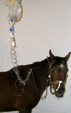
Treatment is supportive only, as there is no antiviral therapy for flaviruses. NSAIDs, such a flunixin meglumine, can be administered to help combat muscle fasciculations. Fluids should be administered to replace water and electrolytes losses. The animal should also receive assisted enteral nutrition via a nasogastric tube until swallowing capabilities return.
 |
| Photo from ckequine.com |
Follow Up
Lucky was showing marked improvement three days following the initial visit. She has regained her ability to swallow and is able to have the NG tube removed. Her gait and incoordination has also shown improvement, and you believe that she will not have any long lasting effects. Horses that have survived WNV infection are immune for life.
Resources
1. Reed, Stephen, Warwick Bayly, et al. Equine Internal Medicine. 3rd ed. St. Louis: Saunders, 2010.
2. Sellon, Debra and Maureen Long. Infectious Equine Disease. 1st ed. St. Louis: Saunders, 2007.
3. Subbiah, E. VM 8124 Course Notes, Equine Viruses I. Spring 2012; Lecture 13-15.

I enjoyed the case based study format. Thought having the preview of the signalment and history of a horse and comparing it to a physical exam made a real life connection. Also the thorough explanation of how to differentiate between the differential list. This would be very helpful when considering real cases. Great job on making me feel like I was doing the case work up! Very eye pleasing in format.
ReplyDelete-Erika
I think I will vaccinate my horses now :) The case study was straight-forward and highlighted important and useful aspects of the disease.
ReplyDeleteGreat Case study! I was unaware that WNV is speculated to cause anti- diuretic hormone release. Very thorough work up with differentials, diagnosis procedures, treatment and prevention. I enjoyed the way you had included commentary between the owner!
ReplyDeleteGreat work
- Katherine
good job
ReplyDelete